CT Scanning Protocol
How to Ensure your Medical (CT) Scan will Work for 3D Modeling
CT scanning protocols informs physicians and other clients whether their protocols are suitable for creating 3D models from the imaging datasets. Datasets with information outside of the specifications below are not suitable for diagnostic models.
Learn more about our FDA approved models.
Thank you for taking a moment to review these protocols. The quality of the CT scan is the most important factor in creating accurate case‑specific anatomical models, bolus designs, and patient‑matched surgical plans. Following the recommendations in this protocol directly impacts the precision of the resulting models, guides, templates, and bolus. We recognize the importance of keeping radiation exposure as low as reasonably achievable, so please apply these guidelines as appropriate for your patients. If you have any questions or would like to discuss the protocol before using it for the first time, please email us at [email protected].
- Please use a 3D scanning routine that provides high resolution images as would be suitable for image guided surgery, stereotactic planning or other 3D applications. It may be useful to consult with your CT vendor’s Application Specialist for advice on optimal parameters for your machine that provide the best scan with acceptable radiation dose levels. Scans should not be taken more than 6 months prior to the surgery date.
- Acquire scans at a high spatial resolution. Series should be acquired with thin, contiguous image slices (equivalent thickness and spacing of 1.25 mm or less) and as small a field of view (FOV) as possible while still including the patient’s anatomy of interest.
- Please provide images in the original scanning plane. If software post-processing is performed to reorient or reformat the scan volume, then a series of thin slice images in the original acquisition plane MUST be included.
- Do not use gantry tilt during image acquisition. Images acquired with gantry tilt then post-processed to reorient images are not acceptable (i.e. “take out” tilt) .
- Please ensure that scans are free from motion artifact. Patient must remain completely still through the entire scan. If patient motion occurs, the scan must be restarted. Image distortion from patient motion can severely compromise the accuracy of a model.
- Image artifact caused by metallic implants can obscure anatomy of interest. Please take steps to minimize artifact from the presence of metal.
- Archive the entire study in uncompressed DICOM format on CD-R or DVD for shipping. It is also possible to transfer image data via the internet. Email us at [email protected] if you have questions.
Recommended protocol for medical CT scanners
| Patient Positioning: | Supine |
| Slice Spacing | 1.25 mm or less (equal to slice thickness) |
| Pixel Size: | 0.60 mm or less |
| Field of View: | Set field of view to include entire area of interest |
| Algorithm: (examples) |
GE- Standard (not bone or detail) Siemens- H30s Toshiba- FC20 Philips- B |
| Gantry Tilt: | 0 o |
| Archive Media: | CD or DVD |
| File Type: | DICOM (uncompressed) |
| Series |
Original/Primary/Axial (No reconstruction, reformat, or process data) |
- Please use a 3D scanning routine that provides high resolution images as would be suitable for image guided surgery, stereotactic planning or other 3D applications. It may be useful to consult with your CT vendor’s
Application Specialist for advice on optimal parameters for your machine that provide the best scan with acceptable radiation dose levels. Scans should not be taken more than 1 month prior to the first treatment date. - Acquire scans at a high spatial resolution. Series should be acquired with thin, contiguous image slices (equivalent thickness and spacing of 3 mm or less) and as small a field of view (FOV) as possible while still including the patient’s anatomy of interest.
- Please provide images in the original scanning plane. If software post-processing is performed to reorient or reformat the scan volume, then a series of thin slice images in the original acquisition plane MUST be included.
- If cavities (e.g. inner ear, outer ear, nose, etc.) will be packed during treatment, acquire the scan with tissue-equivalent packing material. Alternatively, contour packing material during the planning process prior to DICOM submission to 3D Systems.
- Do not use gantry tilt during image acquisition. Images acquired with gantry tilt then post-processed to reorient images (i.e. “take out” tilt) are not acceptable.
- Please ensure that scans are free from motion artifact. Patient must remain completely still through the entire scan. If patient motion occurs, the scan must be restarted. Image distortion from patient motion can severely compromise the accuracy of a model.
- Image artifact caused by metallic implants can obscure anatomy of interest. Please take steps to minimize artifact from the presence of metal.
- Archive the entire study in uncompressed DICOM format on CD-R or DVD for shipping. It is also possible to transfer image data via the internet. Email us at [email protected] if you have questions.
| Slice Spacing: | 3 mm or less (equal to slice thickness) |
| kVp: | 90 - 140 |
| Field of View: | Set field of view to include entire area of interest |
| Algorithm: (examples) |
GE- Standard (not bone or detail) Siemens: H30s Toshiba- FC20 Philips- B |
| Gantry Tilt: | 0 o |
| Archive Media: | CD or DVD |
| File Type: | DICOM (uncompressed) |
| Series: | Original/Primary/Axial (no reconstruction, reformat or post process data) |
- Please use a 3D scanning routine that provides high resolution images as would be suitable for image guided surgery, stereotactic planning, or other 3D applications. It may be useful to consult with your CT vendor’s Application Specialist for advice on optimal parameters for your machine that provide the best scan with acceptable radiation dose levels. Scans should not be taken more than 6 months prior to surgery date for patient 21 years of age or older.
- Series should be acquired with thin, contiguous image slices (equivalent thickness of 1.25mm or less) and as small a field of view as possible while still including the patient’s anatomy of interest.
- Please provide images in the original scanning plane. If software post-processing is performed to reorient or reformat the scan volume, then a series of thin slice images in the original acquisition plane must be included.
- Do not use gantry tilt during image acquisition. Images acquired with gantry tilt then post-processed to reorient images are not acceptable.
- Ensure that scans are free from motion artifact. Patient must remain completely still through the entire scan. If patient motion occurs, the scan must be restarted. Image distortion from patient motion can compromise accuracy.
- Image artifact caused by metallic implants can obscure anatomy of interest. Please take steps to minimize metal.
artifacts; including artifact caused by contralateral implants. Elevation of the contralateral limb to reduce metal artifact scattering to the anatomy of interest is preferred. - Archive the entire study in uncompressed DICOM format on CD-R or DVD for shipping. It is also possible to transfer image data via the internet. Email us at [email protected] if you have questions.
| Patient Positioning: | Position patient in a comfortable, stabilized supine position. Other patient positions are allowed as needed for patient comfort, complete field of view, and minimization of radiation dose. |
| Slice Spacing: | 1.25 mm or less (equal to slice thickness) |
| Matrix: | 512 X 512 |
| kVp: | 90 - 140 |
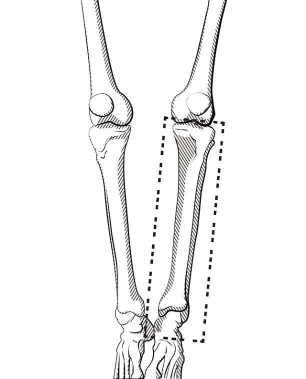
| Field of View: | Capture entire bony segment of interest from joint to joint using smallest field of view possible. See examples below. |
| Algorithm: (examples) | Standard soft tissue reconstruction algorithm preferred. GE: Standard (not bone or detail) Siemens: H30s Toshiba: FC20 Philips: B |
| Gantry Tilt: |
Pelvic Field: 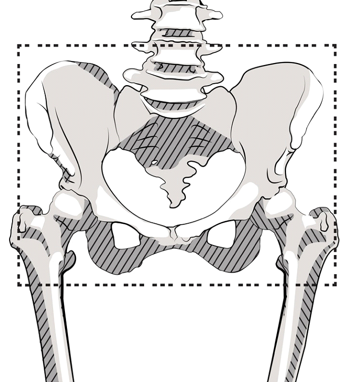
Femoral Field: 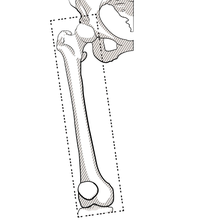
Tibial Field: